Pragniemy poinformować, że filmy instruktażowe są już dostępne
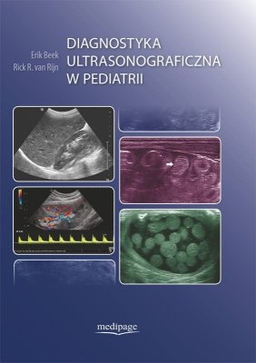
Diagnostyka ultrasonograficzna w pediatrii to książka o przejrzystym układzie z duża liczbą zdjęć z badan USG. Jej przemyślana treść jest zbudowana tak, by być przewodnikiem i odniesieniem w codziennej pracy. Jednoznacznie pokazuje, że badanie ultrasonograficzne winno być pierwszym z wyboru w diagnostyce obrazowej w pediatrii z uwagi na nieinwazyjność, bezpieczeństwo oraz wysoką skuteczność diagnostyczną. Zawiera liczne obrazy prawidłowo zbudowanych narządów i porównuje je ze zmienionymi chorobowo. Książka ta jest przewodnikiem dla każdego lekarza wykonującego badania ultrasonograficzne dzieci.
Zalety książki
- Plan rozdziałów podporządkowany dydaktyce: wprowadzenie, wskazania do badania ultrasonograficznego, technika badania, anatomia prawidłowa z omówieniem wariantów budowy, pomiary z uwzględnieniem wieku dziecka, opis zmian patologicznych każdego rejonu ciała i narządu
- Ponad 2000 wysokiej jakości zdjęć ilustrujących w szczegółach omawiane zagadnienie
- Wyjątkowe „wskazówki od profesjonalistów” zawierające praktyczne porady oparte na doświadczeniu klinicznym zdobytym w pracy z pacjentami pediatrycznymi
- Obszerne piśmiennictwo umożliwiające uzupełnienie treści zawartych w książce
- Filmy instruktażowe - dostępne poniżej (nie wymagają użycia kodów aktywacyjnych)
Diagnostyka ultrasonograficzna w pediatrii to opracowanie niezbędne w pogłębianiu wiedzy i umiejętności wszystkich radiologów pediatrycznych i ogólnych, także początkujących, oraz pediatrów i innych specjalistów, którzy korzystają z ultrasonografii w opiece nad tą wrażliwą populacją, jaką są dzieci.
FILMY WIDEO
Video 3.94 Doppler ultrasound shows flow in the bridging veins
Video 6.12.1 Left aortic arch with aberrant right subclavian artery
Video 6.12.2 Left aortic arch with aberrant right subclavian artery
Video 6.12.3 Left aortic arch with aberrant right subclavian artery
Video 6.13.1a Right aortic arch with aberrant left subclavian artery
Video 6.13.1b Right aortic arch with aberrant left subclavian artery
Video 6.13.2a Right aortic arch with aberrant left subclavian artery
Video 6.13.2b Right aortic arch with aberrant left subclavian artery
Video 6.14.1 Double aortic arch
Video 6.14.2 Double aortic arch
Video 6.14.3 Double aortic arch
Video 6.14.4 Double aortic arch
Video 6.14.5 Double aortic arch
Video 6.14.6 Double aortic arch
Video 6.15.1 Pulmonary artery sling
Video 6.15.2 Pulmonary artery sling
Video 6.1.1a Normal thymus
Video 6.1.1b Normal thymus
Video 6.1.2 Trans-sternal transverse scan
Video 6.1.3 Normal anatomy level of aortic arch
Video 6.1.4 Normal esophagus
Video 6.2 Cervical extension of normal thymus
Video 6.23.1a Thrombus around central venous catheter in the right atrium
Video 6.23.1b Thrombus around central venous catheter in the right atrium
Video 6.23.2 Fibrin sheath of a catheter left behind in the superior vena cava after removal of a central venous line
Video 6.23.3 Embolization of a broken catheter fragment into the
Video 6.4.1 The hemangioma compresses the tracheal lumen to a small gap (arrowheads)
Video 6.4.2 Color Doppler shows the high vascularity and the involvement of the adjacent soft tissues (i.e., thyroid)
Video 6.6.1 The suction tube (arrows) lies in the nondistended proximal pouch (arrowheads)
Video 6.6.2 The distal esophagus is shown behind the heart
Video 6.7 H-type tracheoesophageal fistula
Video 6.8 Achalasia
Video 7.8 Normal air containing lung
Video 7.15 Normal movement of the diaphragm, M mode
Video 7.17a A forked rib
Video 7.17b A forked rib
Video 7.18 Prominent cartilaginous rib
Video 7.33 Thoracic venous malformation
Video 7.40 Pleural fluid with thick echogenic strands after liver biopsy
Video 7.42a Pneumonia complicated by empyema
Video 7.42b Follow up shows a subpleural collection with a thick wall and debris
Video 7.47a A 2-week-old premature with on chest x-ray a persistent opacification in the left upper lung. US shows hyperechoic tissue containing vascular structures
Video 7.47b No air is visible in the lobe. The lung tissue resembles liver tissue
Video 8.6 Video clip shows the IVC located on the left side of the aorta
Video 8.10 Video clip shows the hypertrophied collateral vein leading to the retroperitoneal space
Video 8.16 Doppler US shows an extremely slow flow in the portal vein
Video 8.18 Video shows flow within the metastatic mass and flow within the ascites during respiration
Video 8.20 On respiration flow is visible within the ascites
Video 8.24 Perforation of the gallbladder
Video 8.26 Video clip shows flow of pus within the abscess. Note the deep extend of the abscess
Video 8.27 Video clip shows the flow of pus within the abscess upon compression. Note the rigidity of the surrounding infiltrated fat tissue
Video 8.28 Upon compression flow is seen within the purulent fluid surrounding the small bowel loops
Video 8.31 Free air within the peritoneal cavity between the liver and the abdominal wall
Video 8.35a Video shows independent motion of the tumour in respect to the liver during respiration. This proves that there is no relation between these two structures
Video 8.35b Video shows the extent to the tumour on an axial T2 weighted MRI
Video 8.38 Video shows the extent to the tumour on an axial T2 weighted MRI
Video 8.42 Video shows the extent to the tumour on an axial T2 weighted MRI
Video 8.49 During respiration there clearly is no relation between the cystic mass and the ovary
Video 9.4 Thrombus formation in the umbilical vein
Video 9.16 Choledochal cyst
Video 9.21 Thick mucoid pus within the liver abscess
Video 9.74 Mesenchymal hamartoma
Video 9.79 Hepatoblastoma
Video 9.85 Hepatoblastoma
Video 9.90 Neuroblastoma with encasement of the abdominal vessels
Video 9.98 Tumour in the liver hilum
Video 9.99 US shows air in the portal system
Video 10.32 Splenic lymphoma
Video 10.6 Air artefact from the lung obscuring the spleen
Video 11.2 Juvenile polyp in descending colon
Video 11.4 Patient with mesenteric Burkitt lymphoma with infiltrative invasion of the mesentery
Video 11.5 Esophageal atresia without a tracheoesophageal fistula
Video 11.9 Boy with acute abdominal distention and vomiting
Video 11.11 Hypertrophic pyloric stenosis
Video 11.12 Acute lymphatic leukemia with massive nonstratified wall thickening of the stomach
Video 11.13a Hypertrophic pyloric stenosis
Video 11.13b Normal pylorus
Video 11.14 Normal anatomical position of the D3 segment of the duodenum
Video 11.17 Infant with malrotation and midgut volvulus, whirlpool sign
Video 11.21a Crohn's disease of the terminal ileum
Video 11.21b Crohn's disease of the terminal ileum
Video 11.24 Lobulated character of the polyp and the vessels in the stalk
Video 11.25 Food particles simulating polyps or duplication cysts
Video 11.26a Henoch Schönlein purpura
Video 11.26b Henoch Schönlein purpura
Video 11.28 Meckel's diverticulum
Video 11.30 Necrotizing enterocolitis and intestinal pneumatosis
Video 11.31 Portal vein gas in Hirschsprung's disease
Video 11.32 Pneumoperitoneum in necrotising enterocolitis
Video 11.33 Sloughing of the mucosa in a premature infant with transient ischemia
Video 11.35 Duplication cyst of the ileum in a newborn child
Video 11.37 Postsurgical resolving hematoma or haemorrhagic seroma resembling a duplication cyst
Video 11.38 Benign small bowel intussusceptions
Video 11.39 Extremely large benign small bowel intussusception
Video 11.46 Appendicitis
Video 11.48 Hydropic gangrenous appendix with a torsion at its base
Video 11.53 Neutropenic colitis
Video 11.54 Pseudomembranouis colitis
Video 11.55 Pseudomembranous colitis
Video 11.57 Hemolytic uremic syndrome
Video 11.58 Juvenile polyp in the descending colon
Video 11.59 Encrusted pellets of stools in the colon
Video 11.60 Normal appendage of the colon
Video 11.64 A newborn with a bucket handle deformity of the anus
Video 11.65 Jejunal atresia
Video 11.68 Newborn with cystic fibrosis and meconium ileus
Video 11.72 Meconium peri-orchitis
Video 16.12b Inguinal hernia with a bowel loop
Video 16.12c Inguinal hernia with omentum
Video 16.19 Benign monodermal teratoma
Video 16.29 Torsioned spermatic cord
Video 16.47 Varicocele
Video 16.54a Non-palpable testicles
Video 16.6 Communicating hydrocele
Video 16.7 Non-communicating hydrocele


